Manager Yuan:18018037702
Manager Ma:17705182284
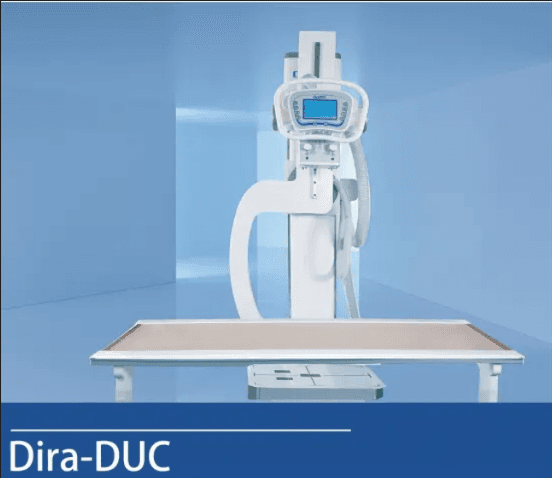
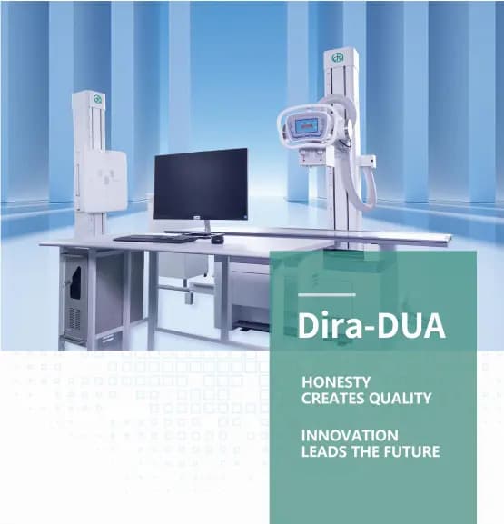
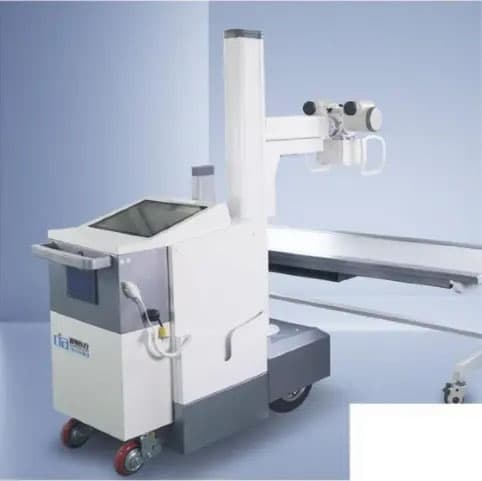
I. Core Imaging System This system adopts a 43×43 cm amorphous silicon flat-panel detector with 9 million pixels (3072×3072 matrix), 139 μm pixel size and 16-bit high dynamic range resolution, ensuring high-quality image acquisition. The imaging time is less than 4 seconds and the spatial resolution reaches 3.5 lp/mm, which meets the requirements for precise clinical diagnosis. II. Intelligent U-C Arm Gantry It delivers flexible positioning performance. The vertical lifting range of the arm is 400–1600 mm, and the moving range of the X-ray tube is 1000–1800 mm. The arm rotates from 0° to 90°, the X-ray tube assembly rotates ±45°, and the detector rotates ±30°, enabling multi-angle radiography. III. Mobile Exam Table Overall dimensions: 2020×660×670 mm. Equipped with an acrylic tabletop, the table bears a load of no less than 100 kg. A mechanical locking device is fitted to ensure stability and safety during examinations. IV. High-Voltage Generator and X-ray Source The 50 kW high-voltage generator supports a maximum tube voltage of 150 kV and an output of 630 mAs. It is equipped with a dual-focus X-ray tube (0.6 mm / 1.2 mm) with an operating tube voltage range of 40–150 kV. The anode heat capacity is 230 kHU, and the housing heat capacity is 1350 kJ (fanless design). The rotating anode speed is 3000 rpm. V. Auxiliary Imaging Components Consists of a vibrating grid and a manual LED collimator. The collimator is powered by 24 V with a power rating of 20 W, featuring 2 mm Al equivalent inherent filtration. The maximum field of view is 48×48 cm (@100 cm SID), and the illuminance is greater than 160 LUX. VI. Acquisition Workstation Configured with Intel i5 processor, 8 GB RAM and 1 TB hard drive. It is equipped with a graphics card of no less than 1600×1200 resolution and a Gigabit Ethernet adapter. The 23-inch color monitor complies with DICOM 3.0 standard for standardized image management. VII. Intelligent Software Functions Remote access to RIS information via Worklist protocol; support for manual registration and emergency quick registration Simple, advanced and custom data query modes DICOM standard disc burning and cross-platform image viewing Query and management of historical images Automatic disk space detection and junk data cleanup Seamless connection with hospital PACS Graphic report system with built-in knowledge base Print patient information and display exposure times on the list Convenient access to original image data